Specular Microscopy
₱ 1,674.00
Specular Microscopy
Specular microscopy is used in measuring endothelial cell density. It is particularly important in :
- Cataract Surgery and Premium IOLs
- Refractive Surgery
- General Corneal Health Assessment
- Contact Lenses
This package is for both eyes.
1,339.20 for Senior/PWD.
Location: St. Luke’s Medical Center Global City Tan Eng Gee Eye Institute
Specular Microscopy
Specular microscopy is used in measuring endothelial cell density. It is particularly important in :
-
- Cataract Surgery and Premium IOLs
Low endothelial cell counts and pre-existing dystrophies can markedly reduce the potential for positive surgical outcomes from an otherwise uneventful cataract surgery.
-
- Refractive Surgery
It is incumbent on refractive surgeons to carefully assess the corneal health prior to performing or recommending an elective corneal surgery to enhance the potential for positive surgical outcomes.
-
- General Corneal Health Assessment
Specular microscopy is an invaluable tool to screen for corneal diseases such as Fuchs’ Dystrophy, keratoconus, other corneal dystrophies, and trauma.
-
- Contact Lenses
Specular microscopy provides a highly detailed view of the integrity and distress of the cornea from the endothelium.
Related products
-
Anterior Segment Photography
₱ 900.00Original price was: ₱ 900.00.₱ 560.00Current price is: ₱ 560.00. -
Biometry
₱ 1,290.00 -
Anterior Chamber Paracentesis
₱ 17,500.00Original price was: ₱ 17,500.00.₱ 12,500.00Current price is: ₱ 12,500.00. -
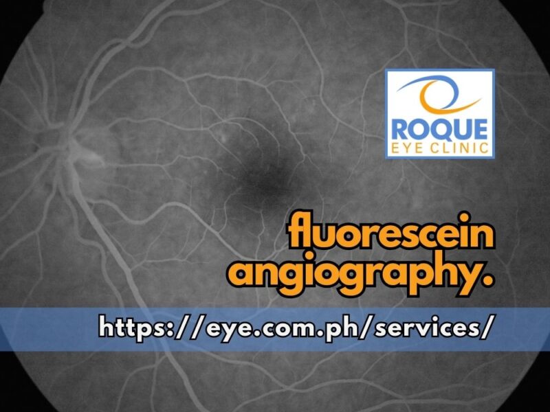
Fluorescein Angiography
₱ 5,250.00 -
Dry Eye Screening Package
₱ 3,936.00 -
Meibomian Gland Expression
₱ 560.00


