
Nystagmus in Children
The term nystagmus is used to describe an involuntary rhythmic movement or oscillations of the eyes. These wobbly eye movements can be characterized as either pendular or jerky. The pendular type is present when the movements have equal speed in each direction. On the other hand, jerk nystagmus is present when a slow movement in one direction is followed by a fast movement in the opposite direction. Nystagmus may also be described as horizontal, vertical or rotatory.
In most cases there is a position of gaze where the nystagmus diminishes and this is referred to as the null point. Children with nystagmus often adopt a compensatory head turn to maintain both eyes at the null point, thereby improving visual acuity. A comprehensive eye examination by the pediatric ophthalmologist can help identify good candidates for nystagmus surgery (Kestenbaum-Andersen procedure) as well as identify other associated problems.
It is helpful to classify nystagmus as either congenital (present at birth or within months after birth) or acquired.
As the name implies, the onset of congenital nystagmus is early, usually the first few months of life. Because of this early onset, the brain is able to suppress the motion. Therefore these infants do not perceive the cyclic wobbly eye motion associated with nystagmus. There are 2 basic subtypes: congenital motor, and sensory.
Congenital motor nystagmus (infantile nystagmus) is usually present in both eyes and symmetric. It occurs during the first few months of life and is often inherited as an x-linked trait (FRMD7 gene). The compensatory head turn that minimizes the nystagmus is usually established by 2-4 months of age, or as soon as the child gains head and neck control. These children have relatively good visual potential (usually around 20/50 or better), particularly when they assume the compensatory head turn.
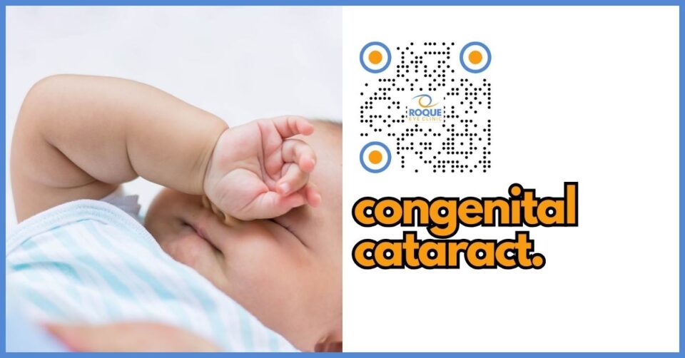
Congenital sensory nystagmus is due to the lack of the fixation reflex secondary to neonatal blindness. Any disease that results in bilateral neonatal blindness such as congenital cataract, corneal opacities, congenital optic nerve atrophy or hypoplasia, and congenital retinal disorders, can cause this form of congenital nystagmus. The pattern of sensory nystagmus is usually indistinguishable from congenital motor nystagmus, except that the nystagmus has a larger amplitude and the movements show poor fixation with a searching character. The onset is later than that seen in congenital motor. Patients with sensory nystagmus rarely adopt a compensatory face turn because their visual potential is not very good despite changing head position.
Acquired nystagmus, may be a sign of a serious neurological condition, and therefore warrants a neurology consult. Neurologic disease involving any part of the brain can cause nystagmus and is often associated with the perception of the environment moving, or oscillopsia. Only patients with acquired nystagmus will experience oscillopsia; as these patients do not have the neural plasticity to suppress the shaking image. Oscillopsia, therefore, is an important indication that the nystagmus is acquired.
A comprehensive eye examination by the pediatric ophthalmologist can help identify good candidates for nystagmus surgery, as well as identify other associated problems. Andersen procedure requires surgery on two yoke muscle, one from each eye, for the mild to moderate head turns. Kestenbaum-Andersen procedure, on the other hand is recommended for large head turns, requiring surgery on four horizontal rectus muscles. These procedures adjust the position of the eyeballs to the primary position and gets rid of the head turn. Patients with significant refractive errors are expected to need glasses even after surgery.
- Graf, M., Hausmann, A. and Lorenz, B. (2019). High-dose Andersen operation nystagmus-related anomalous head turn. Graefes Arch Clin Exp Ophthalmol, 257(9): 2033-2041.
- Ospina, L. (2018). Dealing with nystagmus. J Binocul Vis Ocul Mobil, 68(4): 99-109.
- Papageorgiou, E., McLean, R., and Gottlob, I. (2014). Nystagmus in childhood. Pediatr Neonatol, 55(5): 341-351.
