
REFRACTIVE SURGERY SCREENING INSTRUCTIONS
- CONTACT LENS HOLIDAY. Soft contact lens (SCL) users are required to discontinue the use of SCLs for at least 1 week prior to the refractive screening. Hard contact lens and Rigid Gas Permeable (RGP) users are required to discontinue the use of RGPs for at least 4 weeks prior to the refractive screening. Contact lens holidays allow the surface of the cornea to recover to its natural state, thereby increasing the reliability of the results of the diagnostic screening. Kindly wear your prescription eye glasses during this contact lens holiday. Please coordinate closely with our staff if you wish to move the tests to an earlier or later date.
- OCULAR LUBRICATION. Kindly apply over-the-counter preservative-free lubricating drops hourly to both eyes during waking hours for at least 1 week prior to the surgery. This will make you feel comfortable with instilling topical medication to your eyes after surgery.
After complying with your refractive surgery pre-screening instructions, you are now ready to proceed with your refractive surgery screening. It usually takes an average of three (3) hours to complete a refractive surgery screening. Your eyes will remain dilated for 6-8 hours after the procedure, therefore it is recommended that you don't schedule anything important for the rest of the day.
After reporting to our clinic, our staff will escort you to the laser room where the process will begin.
DIAGNOSTICS
PERSONAL INFORMATION.
Your personal information will be logged into the hospital records.
CLINICAL HISTORY.
Clinical history taking will be performed. Your ophthalmic history, medical history, and family history will be reviewed. Please take note of all the medications that you rare currently taking.
PRESCRIPTION GLASSES.
Please hand over all of your current prescription eye glasses so that our optometrist may record the power.
VISION AND REFRACTION.
Your visual acuity will be taken with and without your current prescription eye glasses. Automated refraction, manual/manifest refraction and dilated/cycloplegic refraction will be done. All these measurements are critical in determining your best potential visual acuity.
SLIT LAMP BIOMICROSCOPY.
Your eyelids, eyelashes, and the anterior segment (conjunctiva, cornea, pupil, iris, and lens) of your eye will be examined with a slit beam of light. This examination is performed to search for lid and lash conditions, corneal diseases, pupil and iris abnormalities, and lens status.APPLANATION TONOMETRY (Haag Streit Goldmann).
A topical anesthetic, proparacaine, will be instilled into your lower eye lid pocket in order to numb your eye. A fluorescein dye will be used together with a cobalt blue filter to measure your ocular pressure. Your eye pressure will be correlated with your corneal thickness. This examination is used to screen for glaucoma.
OCULAR SURFACE EVALUATION.
Fluorescein dye applied on your eye will be used to examine the ocular surface for signs of dry eye disease. Your tear break up time (TBUT) will be measured. In some instances, the use of a Rose Bengal or Lissamine Green dye strip may be necessary.
PUPILLOMETRY (Oasis Medical Colvard).
An infrared pupillometer will be used in a dark room to check for your pupil size at night. This will assist us in selecting an appropriate optical zone for best night vision. This examination is performed prior to pupil dilation.
OPTICAL BIOMETRY (Zeiss IOL Master or Haag Streit Lenstar).
Your ophthalmic 'time capsule' will be taken to secure your pre-refractive surgery corneal curvature and eyeball length. This will be useful once you have cataract surgery in the future. This examination is performed prior to pupil dilation.
SPECULAR MICROSCOPY (Konan or Topcon).
This measures endothelial cell density (ECD). Low ECD is a risk factor for refractive (corneal and cataract) surgeries that can be missed without the use of a specular microscope. This examination is performed prior to pupil dilation.
CORNEAL TOPOGRAPHY (Zeiss Humphrey Atlas or Orbscan).
The corneal curvature is measured using this diagnostic test. It will show the flat and steep areas of your cornea. This helps in screening for keratoconus, which is a contraindication to laser refractive laser surgery. This examination is performed prior to pupil dilation.
SCHEIMPFLUG ANALYZER (Oculus Pentacam or Zeimer Galilei).
This is an improved version of a corneal topographer. It has the benefit of giving refractive surgeons more information on the curvature and thickness of the cornea. It is very useful in identifying even early keratoconus. This examination is performed prior to pupil dilation.
ABERROMETER (Zeiss WASCA or AMO Wavescan).
Think of this as a super automated refraction machine. Traditionally our refractive errors are determined using an auto refractor, phoropter, or trial lenses. These are able to determine the amount of myopia, hyperopia and astigmatism. However, our eyes as an optical system have higher wavefront measurements that are of consequence to the quality of our vision. The wavefront aberrometer measures the way a wavefront of light passes through the cornea and the crystalline lens, which are refractive components of the eye. Distortions that occur as light travels through the eye are called aberrations, representing specific vision errors. This examination is performed before and after pupil dilation.
OCT MACULA (Zeiss Cirrus OCT or OptoVue OCT).
A special photo of the macula is taken with this machine. The macula is responsible for the central 30 degrees of functional vision. So a fully functioning macula will allow us to see the face of an individual who we are looking at. If this is diseased, we will see a central obstruction in vision. This examination is performed before or after pupil dilation.
OCT OPTIC NERVE (Zeiss Cirrus OCT or OptoVue OCT).
A special photo of the optic nerve is taken with this machine. The optic nerve may be damaged because of glaucoma. Early detection is possible with this diagnostic test. This examination is performed before or after pupil dilation.
DILATED FUNDUS EXAMINATION.
Dilating ophthalmic drops will be instilled into your lower eye lid to make your pupils dilate. It usually takes around 30 minutes to achieve full pharmacologic pupillary dilation. Dilation will allow us to examine your retina for congenital/acquired posterior segment diseases. We can see retinal tears/holes, macular degeneration, optic nerve diseases, among other things. The presence of any posterior segment condition may disqualify one from having refractive laser surgery. This is performed by Dr. Roque at the end of the refractive surgery screening process.
REFRACTIVE COUNSELLING.
Dr. Roque will evaluate the results of your refractive surgery screening and determine if your eyes are fit to undergo refractive surgery. It is at this time that you will be able to ask all your questions about the possible surgical options. If you are accompanied by a decision maker prior to surgery, you may have this person join you for this session.
LEARN MORE ABOUT REFRACTIVE SURGERY

BOOK AN APPOINTMENT
It takes less than 5 minutes to complete your online booking. Alternatively, you may call our BGC Clinic, or our Alabang Clinic for assistance.
OUR REFRACTIVE SURGERY SPECIALIST

DR. MANOLETTE ROQUE
MD, MBA, DPBO, FPAO, FPCS
Dr. Manolette Roque is a specialist in uveitis, cataract, and refractive surgery. His private practice began in 2000, after his post-graduate fellowship at the Massachusetts Eye and Ear Infirmary, Harvard Medical School, in Boston, Massachusetts, USA. His patients are mostly adults who desire spectacle independence. His advocacy includes taking care of individuals with ocular inflammatory diseases.
Everyone deserves the best eye care possible.
OUR CLINICS
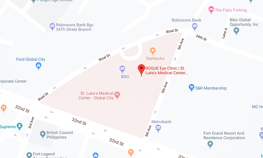
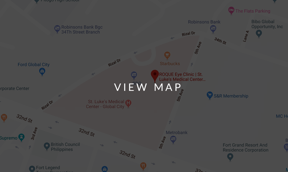
BGC CLINIC
- ST. LUKE'S MEDICAL CENTER GLOBAL CITY
2/F Medical Arts Building 217
Rizal Drive corner 5th Avenue
Bonifacio Global City, Taguig 1634
Philippines
SLMC CLINIC HOURS
- 9am - 12pm
Appointments only
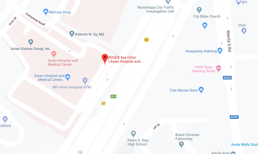
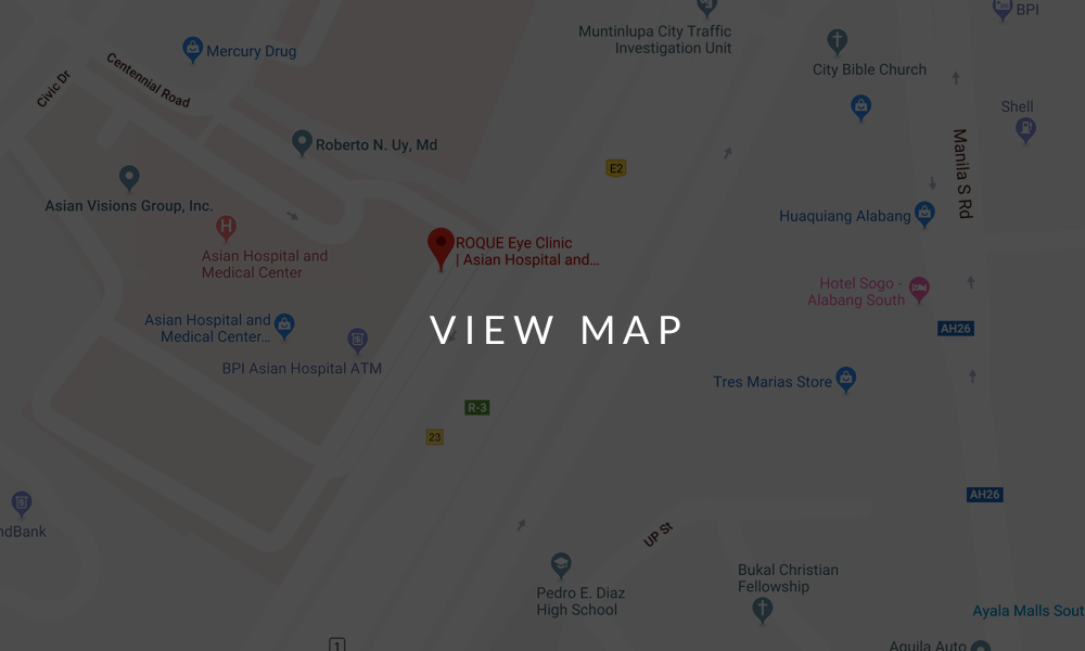
ALABANG CLINIC
- ASIAN HOSPITAL AND MEDICAL CENTER
5/F Medical Office Building 509
2205 Civic Drive, Filinvest City
Alabang, Muntinlupa 1781
Philippines
AHMC CLINIC HOURS
- 1pm - 4pm
Appointments only
BOOK AN APPOINTMENT
It takes less than 5 minutes to complete your online booking. Alternatively, you may call our BGC Clinic, or our Alabang Clinic for assistance.


