Normal Intraocular Pressure
One of the main risk factors for the development of glaucoma is a relatively increased intraocular pressure (IOP). The normal level of IOP varies from person to person. The statistically “normal” IOP is 16.5 mmHg (millimeters of mercury) with pressures above 20 mmHg considered statistically “elevated”. These values were obtained by averaging the IOP results of thousands of normal individuals with a wide range of pressures (from 10 to 35). For example, if a person with a usual IOP of 9 mmHg were to later have an IOP of 19 mmHg that would be a significant elevation even though it’s still a statistically normal IOP. Thus, a person may have an IOP that is increased relative to their normal IOP but is still not increased compared to the rest of the population (absolute increase). Ophthalmologists always keep this in mind when making the diagnosis of glaucoma and deciding what IOP to aim for when treating glaucoma patients.
Intraocular Pressure Fluctuation
The actual level of IOP is not the only aspect of IOP that matters in glaucoma. Our IOP normally fluctuates throughout the day. Glaucoma patients tend to have a wider daily fluctuation in their IOP (difference between peak IOP and trough IOP). This is called the diurnal variation. The IOP reading taken at the doctor’s office is the measurement for that specific moment only and does not indicate what the IOP levels have been the rest of the day. This is why glaucoma patients or glaucoma suspects are sometimes asked to have several IOP measurements taken in a single day. This is procedure is called phasing or diurnals.
Intraocular Pressure Lowering
Intraocular pressure is not the only risk factor for glaucoma but it is the one that ophthalmologists (and some glaucoma patients, too) are most preoccupied with. This is because IOP is the only risk factor that we are able to modify. We have many drugs and treatments that can lower IOP but there are no treatments (yet!) that can decrease the effects of aging or that can change a person’s genetic make-up. (Indeed, gene therapy may already be available for certain diseases but, unfortunately, research has not yet even uncovered all of the genes that predispose to the various types of glaucoma.) Evidence has shown that lowering intraocular pressure and decreasing intraocular pressure fluctuation are effective for preventing glaucoma progression.
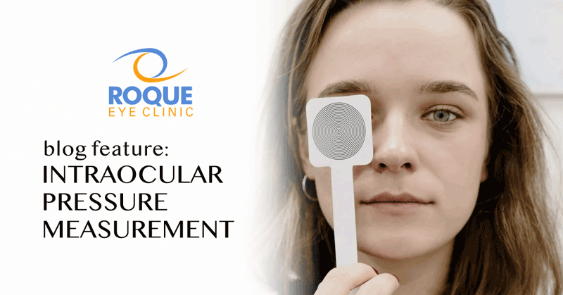
Intraocular Pressure Measurement
Intraocular pressure can be measured using various instruments. The current gold standard instrument for measuring IOP is the Goldmann applanation tonometer (GAT or AT). It is called the gold standard because it gives the most accurate, reliable, and reproducible measurements. It measures IOP by flattening an area of the cornea and measuring the amount of pressure needed to flatten that area of the cornea. Because the instrument needs to come into contact with the eye your doctor will first put anesthetic drops to numb your eye and fluorescein dye drops to make your tear film more visible during the measurement process. The other instruments for measuring IOP work in different ways and have varying degrees of accuracy. There are promising new IOP-measuring instruments being studied and being introduced into the market and one of those instruments may one day replace the AT as the gold standard for measuring IOP.
Central Corneal Thickness and Pachymetry
Most methods of IOP measurement including AT can be affected by corneal thickness. An unusually thick or thin cornea can cause the IOP measurements to be erroneously high or erroneously low, respectively. A central corneal thickness outside if the normal range has also been associated with a difference in the risk of glaucoma or glaucoma progression. Those with thinner corneas seem to have a slightly higher risk of having glaucoma. If your doctor suspects that your corneal thickness will have a role in the management of your glaucoma you may be asked have your corneal thickness measured. This simple test is called pachymetry and there are different kinds of instruments that can be used. The most commonly available method involves lightly touching an ultrasonic probe to the anesthetized cornea for a few seconds.
References:
- Kahn HA et al. The Framingham eye study. 1. Outline and major prevalence findings. American Journal of Epidemiology 1977; 106:17.
- Heijl et al. Reduction of intraocular pressure and glaucoma progression: Results from the Early Manifest Glaucoma Trial (EMGT). Archives of Ophthalmology 2003; 121:48-56.
- Asrani et al. Large diurnal fluctuations in intraocular pressure are an independent risk factor in patients with glaucoma. Journal of Glaucoma 2000; 9:134-142.
- The AGIS Investigators. The Advanced Glaucoma Intervention Study (AGIS): 7. The relationship between control of intraocular pressure and visual field deterioration. American Journal of Ophthalmology 2000; 130:429-440.
- Collaborative Normal Tension Glaucoma Study Group. Comparison of glaucomatous progression between untreated patients with normal tension glaucoma and patients with therapeutically reduced intraocular pressures. American Journal of Ophthalmology 1998; 126:487-97.
- Kass et al. The Ocular Hypertension Treatment Study: A randomized trial determines that topical ocular hypotensive medication delays or prevents the onset of primary open-angle glaucoma. Archives of Ophthalmology 2002; 120:701-713.
- Ritch R, Shields MB, Krupin T (Eds). The Glaucomas, 2nd Edition. St. Louis, Missouri, USA, 1996, Mosby-Year Book, Inc.
- Epstein DL, Allingham RR, Schuman JS (Eds). Chandler and Grant’s Glaucoma, 4th Edition. Baltimore, Maryland, USA, 1997, Williams & Wilkins.